


Have you ever had knee pain? It is extremely uncomfortable when you suddenly start limping or can’t go down to the ground without pain in your knees. Arthrosis of the knee joint is not life-threatening, but it dramatically degrades its quality.
What is knee arthrosis?
Knee arthrosis(gonarthrosis, osteoarthritis, knee joint osteoarthritis). Gonarthrosis is arthrosis of the knee joint (this disease has nothing to do with gonorrhea). In advanced cases, nothing but surgery helps. You need it? Then don’t run into that state.
Causes of knee arthrosis.Distinguish between primary and secondary osteoarthritis of the knee joint. If the cause of the disease is not revealed, such arthrosis is called primary, inherited through the maternal lineage. If the grandmother suffers from arthrosis of the knee joints, this disease can also occur at a younger age for her daughter and granddaughter.
Secondary arthrosis is caused by trauma, congenital disorders of the knee joint, physical overload (sports, occupation), endocrine disorders.
Risk factors are overweight, female gender, old age. Cartilage is very sensitive to a decrease in female sex hormones, during menopause all joints begin to "collapse. "Therefore, overweight older women are more severely and more likely to suffer from knee arthrosis.
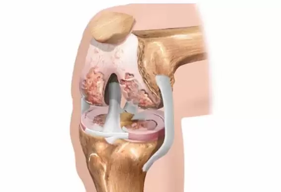
Knee anatomy.The knee joint is made up of the femur, tibia and patella. The joint surfaces of the bones are covered with a layer of cartilage. Extra cartilaginous spacers between the bones are called meniscus and are padded. The knee joint has the largest synovium, which forms large twists and bursaes.
The joint cavity is filled with joint fluid that feeds the articular cartilage. The synovial fluid contains hyaluronic acid, which is needed for even sliding of the joint surfaces. The ligaments, muscles and tendons control and restrict joint movement.
General description.With arthrosis of the knee joint, the articular cartilage is destroyed. Knee arthrosis has three stages. In the first stage, the articular cartilage and menisci feed is interrupted. Cartilage loses its elasticity and ruptures. Abnormal friction occurs between the bones. Joint overload is accompanied by inflammation and pain in the knee.
In the second stage, the destruction of articular cartilage and the meniscus begins. The bone responds to the load with marginal increases - osteophytes ("thorns"). The amount of intra-articular fluid decreases and the narrowing of the joint space increases. As a result, knee pain occurs during normal exercise, walking.
In the third stage, the pronounced bone deformity of the knee joint appears with a sharp restriction of natural movements.
Symptoms of knee arthrosis.The main symptoms of arthrosis are pain, limited mobility and deformity of the knee joints. Arthritis of the knee joint is long-term, with symptoms slowly, irreversibly increasing. If knee pain suddenly, suddenly, first appeared, it is probably not arthrosis.
Arthrosis of the knee joint gradually begins with discomfort or less pain in the knee during overload, long walking as they go down, rising from a squatting position. At rest, the pain goes away quickly.
In the second stage, knee pain already appears with normal effort. The volume of active movements decreases in the knee joint. The shape of the joint changes due to bone deformity and the accumulation of abnormal fluid.
In the third stage, the pain becomes chronic, not only during movement, but also at rest. Nocturnal pains disrupt sleep. The knee has a hard time getting into bed without pain. Swelling of the joint indicates an increase in inflammation. The mobility of the knee joint is minimized.
The joint is significantly deformed and the legs become O- or X-shaped. In severe cases, complete destruction of the joint results in the development of ankylosis (immobility).
There are 4 types of pain with knee arthrosis:
- the mechanical type of pain occurs as a result of daytime physical activity and subsides during the night rest period. These knee pains are associated with a decrease in the shock-absorbing capacity of cartilage and bone structures. Knee pain is usually localized in the anterior and inner region of the knee joint and in the upper part of the lower leg.
- nocturnal pain is associated with stagnation of venous blood, increased intraosseous pressure in the joint, and inflammation.
- The "onset" pain occurs after a rest period, disappearing 15-20 minutes after movement in the joint. These knee pains are caused by friction on the joint surfaces on which fragments of cartilage breakdown are deposited.
- persistent knee pain is caused by muscle cramps as well as the development of synovitis.
Complications of knee arthrosis.Synovitis is an inflammation of the membrane of the joint cavity that covers the joint cavity from the inside. Signs of inflammation: swelling, fever, redness, pain, joint dysfunction.
Normally, the knee joint contains 3-5 ml of synovial fluid. Diseases in the joints produce increased inflammatory fluid. The amount of effusion (abnormal fluid) can reach 30-70 and even 100 ml. The knee outflow first fills the cavity inside the patella (medial fossa). As the volume increases, the upper volvulus fills, with a massive swelling above the patella ("horse saddle").
Baker cyst occurs with a significant increase in the volume of intraarticular fluid. A round, flexible bulge forms in the popliteal region. It is not a tumor, it is not an oncology, and it does not need surgery. A Baker’s cyst can cause discomfort, pressure, and pain in the knee while moving. The diameter of the cyst is between 2 and 6 cm. At even larger sizes, the cyst can pinch the nearby peroneal nerve by developing weakness and numbness in the foot.
Diagnosis of knee arthrosis.Laboratory tests are not suitable for making a diagnosis, but are intended to rule out other conditions associated with knee pain. In arthrosis, indicators of blood counts are within normal limits of leukocytes and ESR without inflammatory changes. Rheumatic tests are negative. Uric acid levels are within the normal range.
X-rays show bone changes in the joint, ruling out the traumatic causes of joint pain. In Hungary, the X-ray classification of arthrosis is used according to stages.
Stage 1 - the presence of marginal bone growth with a slight narrowing of the joint space;
Stage 2 - the joint space narrows more clearly, subchondral sclerosis occurs;
Stage 3 - sharp narrowing of the joint space, flattening of the joint surfaces, formation of cysts;
Knee MRI is indicated in the early stages of the disease, when radiological changes are not yet visible, but the patient has typical knee pain. With the help of MRI you can assess the condition of cartilage, meniscus, ligaments, tendons. Ultrasound of the knee joint helps to visualize the soft tissues (meniscus, muscles, ligaments) and to assess the volume of effusion.
Arthroscopy is the most accurate method for diagnosing knee arthrosis. A special probe is placed in the joint cavity and the doctor assesses the extent of cartilage destruction under a microscope.
Treatment of knee arthrosispresents a difficult task. In each case, you must choose an individual treatment program.
When you start saying banal things during a consultation, patients look surprised at first. Is that where we came from? Give a wonderful injection so that my knee never hurts again. We need to explain that there is no single method that can eliminate arthrosis. To recover, you need to move, lose weight, sign up for the pool. And a person wants to lie on the couch, grow a "beer belly, " grab the problem with a lot of medicine, and be healthy. But unfortunately !!! In this case, medicine is powerless.
Painkillers do not cure, they only relieve pain. Anti-inflammatory drugs are prescribed only at the time of worsening knee pain. Some non-steroidal medications contribute to the further destruction of cartilage by relieving pain. Healing ointments do not cure knee arthrosis, but relieve knee pain. In case of edema, redness of the joint, warming ointments and compresses are contraindicated; it is better to use topical medications with non-steroidal anti-inflammatory drugs.
Chondroprotectors do not relieve pain, are expensive and need to be taken for a long time. I consider them "dummies" and practically don't designate them. Currently, avocado and soy extracts have appeared in pharmacies, but I have not used this drug in my clinical practice and I have no opinion of its own on its effectiveness.
Appropriate physiotherapy exercises in a sitting or lying position are required to treat and prevent knee arthrosis. Squatting and jumping are strictly forbidden. Cycling, swimming or exercising in the water, skiing is useful. Labor exploitation in the country often causes increased pain in the knee. With arthritis of the knees, running, fast walking on inclines and weight lifting are not recommended.
Diet for arthrosis of the knee joint.The knee joints carry their own load. Therefore, overweight people should lose at least 3-5 kg. Some patients have to lose more than a dozen pounds. Otherwise, no treatment will be effective. It is not necessary to "sit" on some diet, it is harmful to the body.
For the rest of your life, you need to change your eating behavior, just "don't love" all the harmful products (sweet, starchy foods, beer, etc. ). Proper eating should become a habit. To lose weight, you should eat adequate food every 3 hours.
To reduce inflammation in the joints, homeopathies recommend foods that alkalize the blood and intraarticular fluid. To this end, the consumption of meat should be sharply limited and the amount of vegetables and fruits in the diet should be increased.
It is believed that sausages, sausages, smoked meat, fast food enhance the inflammatory processes in the joints. Instead of pharmaceutical chondroprotectors, I suggest eating properly prepared jelly meat.
Orthopedic correction reduces stress on the knee joints. If you have pain in your knee joints, you should pick up your kneecap. In advanced cases, walking with a reed is indicated. A heel insert is recommended for shortening the foot. Recently, it has become fashionable to use kinesis tapes. These are adhesive tapes made of natural cotton that are glued around the affected knee, not limiting its mobility, but helping to relieve the joint and reduce muscle cramps.
I consider interstitial electrical stimulation to be the most effective method of treating pain in arthrosis. In combination with hirudotherapy (leech therapy) and pharmacopuncture, VTES gives very good results. I will give a case from practice.
A 54-year-old, II. A man with stage right knee arthrosis turned to me for help. The knee pains bothered him for 6 years. Over the years, she has received several drug therapy, physiotherapy, corticosteroid blockade treatments, and has attended repeated courses at a rehabilitation center. But the patient's condition only got worse. He came to me for a consultation on whether to undertake joint replacement surgery or try something else conservatively. I didn’t have to persuade him for a long time, he immediately agreed to the treatment I suggested.
In the first session, I gave him 6 leeches to help him cope with joint swelling and eliminate nighttime pains. The knee can be moved more easily and freely. The man was a little relieved. We then performed 3 interstitial electrical stimuli and almost completely stopped the pain syndrome.
Subsequently, the success was consolidated by the introduction of anti-inflammatory and chondroprotective homeopathic remedies into acupuncture points. Three weeks after the start of therapy, the patient dropped the cane and began to move freely without limping. 3 years have passed since then. The knee pains did not return. Once a year, we hold a meeting of the VTES for preventive purposes.
Intra-articular injections with hormones are very effective in relieving severe pain, swelling and inflammation in an emergency. The indication is effusion, it is forbidden to block with corticosteroids in the "dry joint"! They temporarily relieve the pain, but such injections do not cure the arthrosis itself, and the cartilage that follows them is even more destroyed. These should be performed by a specially trained physician who is familiar with the indications, contraindications, medications, and points of application. A total of up to 3 blocks are required per bond.
After removing the swelling and inflammation, hyaluronic acid preparations, so-calledLiquid prostheses are injected into the joint. As a natural lubricant, they act on the joint, improving the slippage of bone surfaces and restoring the shock-absorbing functions of cartilage. But hyaluronic acid preparations are expensive and only last for 6-8 months. There is no point in giving hyaluronic acid preparations with complete loss of joint space and in patients over 65 years of age.
Treatment with folk remedies.You can use tincture or decoction of cinquefoil, compress radish, horseradish or ginger, turpentine baths.
Joint endoprosthesis should only be performed in case of severe dysfunction of the knee joint, because after 10-15 years this joint must be changed again. Will there be enough strength and health every 10-15 years for surgery under general anesthesia and subsequent rehabilitation??? Therefore, do not rush to agree to an operation! Take care of your joints!